Light-sheet microscopy is a powerful tool for 3D imaging of intact living model organisms. It uses a thin sheet of light to excite just the fluorophores within the focal area. This allows them to capture optical sections, including axial resolution (the ability to discern two structures along or parallel to the path of the fluorescent beam).
Because of this setup, light-sheet microscopy has the advantage of limiting the area of the sample subject to photobleaching and phototoxicity, meaning sample integrity remains over longer imaging periods. Light-sheet imaging can also record tens to thousands of images per second. It’s ideal for detailed analyses of large, optically cleared samples, providing high spatio-temporal resolution for live-imaging biological specimens over longer periods.
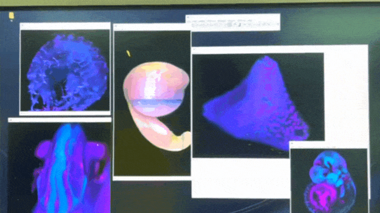
Light-sheet images captured by attendees of the Developmental Biology Course with the support of the LiSIUM team. | Video courtesy of Lovy
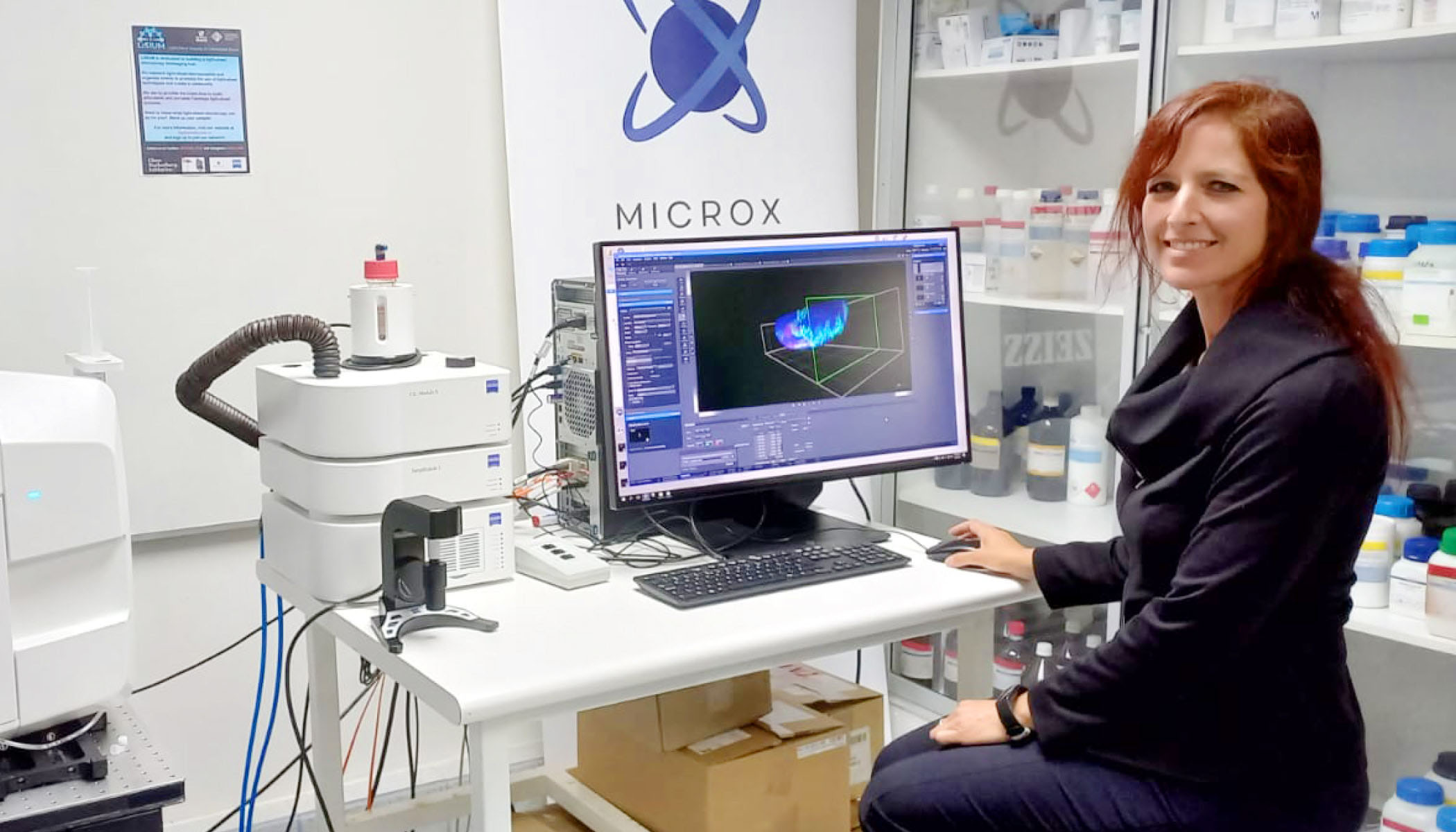
Lovy was eager to try the light-sheet microscope at the Universidad Mayor for her research.
“I was really excited to start to learn more about this technology,” she says. “But our biggest mission is to share the technology with others because it’s the only one of its kind basically on the continent.”
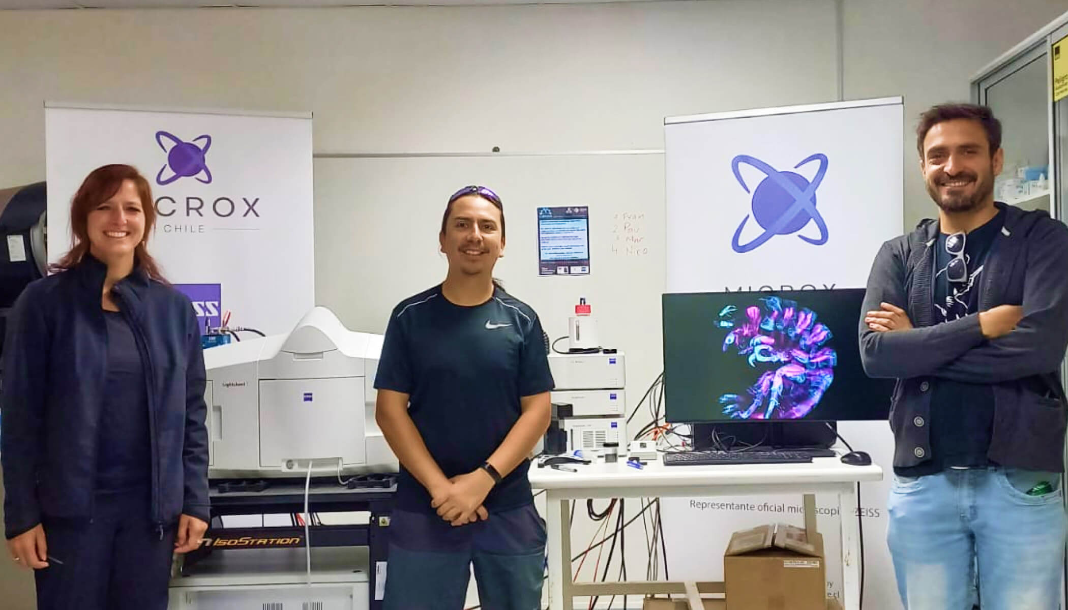
To enable this, Lovy led an Expanding Global Access to Bioimaging grant application alongside Felipe Court and Leonardo Valdivia to begin building networks of researchers called the Light-Sheet Imaging at Universidad Mayor (LiSIUM). LiSIUM connects five labs at Universidad Mayor, five labs in other institutions in Chile, and four international labs in South America to share information about this instrument.
The hub also leverages workshops, conferences and experts to make the technology more accessible for biomedical researchers. For example, Lovy’s team coordinated training opportunities offered concurrently with a world-class European Molecular Biology Organization developmental biology course, bringing together experts in model organisms suitable for light-sheet imaging.
Building sustainable for sustainable access
Once Lovy’s team established a foundational network for light-sheet microscopy, she wanted to find a way to remove additional barriers to accessing this technology. When attending a light-sheet microscopy conference in 2020, Lovy learned about the compact and portable light-sheet microscope developed by Jan Huisken, nicknamed “Flamingo” for its one-legged stand and vertical profile. Not only is the Flamingo light-sheet microscope small enough to fit into a suitcase, but it’s also relatively easy to set up in a new location with the remote configuration offered by Huisken as needed.