Csaba Forró in the lab at Chicago Biohub.
Back to life/science blog
Bioengineering Chicago 8 min read

Building devices that unite cells and sensors

Csaba Forró, a Chicago Biohub group leader, discusses how merging physics, engineering, and biology helps researchers investigate disease at the cellular level.

Csaba Forró in the lab at Chicago Biohub. (Credit: Dale Ramos, CZI)

Collin Blinder August 6, 2025

When you think about biomedical engineering, your brain probably doesn’t jump right to kirigami, the Japanese craft of forming paper sheets into 3D forms. But for biomedical engineer Csaba Forró, kirigami is one of many tools that can be used to study inflammation and disease.

Forró leads the Biomimetronics group at Chan Zuckerberg Biohub Chicago, engineering innovative devices that can seamlessly interface with lab-grown human tissue. The group is creating non-invasive ways to study the human body, from building electrical sensors that detect seizure activity in Petri dish brain cells to developing mechanisms that stretch lab-grown skin. Their next-generation devices can both monitor and manipulate cells, helping Biohub scientists study how cells behave in real time and paving the way for a deeper understanding of health and disease.

Forró’s journey into experimental bioengineering began with a pivot. After studying physics as an undergraduate at EPFL, Switzerland, he pursued a master’s degree in theoretical physics. But Forró soon discovered that his true calling was in applied research.

After six months, I realized I didn’t want to spend a decade just to acquire the fundamentals I need to start contributing to the field," he recalls. "I was eager to build things, test ideas in the lab, and see results unfold in real time.”

Motivated by this drive to make a more immediate impact, Forró transitioned into laboratory research, earning his Ph.D. in neuroengineering at ETH Zürich. There, he developed devices and platforms to physically monitor brain synapses and discovered a passion for combining computer simulations with precision device engineering. He also made contributions at the intersection of artificial intelligence and microscopy, earning him ETH Zürich’s Best Thesis of the Year award and described in a Nature Machine Intelligence paper.

After becoming captivated with applied research, Forró secured the Marie Sklodowska-Curie Global Fellowship, which brought him to Stanford University for postdoctoral research. There, Forró built custom electronics to study lab-grown human brain organoids. He used concepts from kirigami to design two-dimensional sensor arrays which unfurl into three-dimensional structures. These flexible devices cradle and conform to the organic shapes of living tissue, uniting biology and electronics in a natural, non-disruptive way. This innovative approach marked the emergence of a new class of bio-integrative, non-invasive devices capable of monitoring and interacting with cells, as described in a Nature Biotechnology paper.

Now, Forró’s group is building such thin sensors that tissue samples can actually grow around them, allowing researchers to damage the tissue and monitor the ensuing inflammation response. Such in-depth research will help Biohub scientists reach their goal of understanding inflammation at the cellular level, leading to treatments and preventative interventions for inflammatory disease.

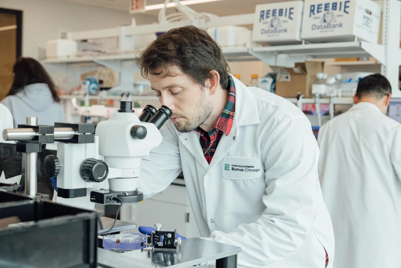
We recently chatted with Forró about his work at the Chicago Biohub and the future of non-invasive biomedical research.

What kinds of research does your group conduct at the Chicago Biohub?

I’ve been particularly interested in the potential of patient-derived stem cell technology, which allows us to reprogram adult cells, like skin or blood cells, into stem cells and then grow virtually any human tissue — such as heart, lung or brain cells — in the lab. This approach lets us recreate disease-specific tissue in Petri dishes and then conduct detailed, real-time studies without needing to perform surgery on humans or work with animal proxies.

Where we come in is figuring out how to study the tissue that’s grown with some sort of disease, such as epilepsy or Parkinson’s. You need to measure the electrical activity, the chemical contents, neurotransmitters, and all their inter-related dynamics. So my group builds instruments that collect data from those tissues. But these devices don’t just record data — we can also stimulate cells or test potential medical interventions directly in human tissue samples. For example, right now we’re building devices for stretching lab-grown skin to help study psoriasis, which is a disease that seems to get exacerbated in regions where the skin stretches a lot.

In what ways do you hope to advance the field of biomedical research?

My postdoctoral research at Stanford was mainly focused on developing devices that could sense electrical signals from neural tissue samples. Now, my group at the Biohub is expanding on that vision significantly. We’re creating platforms that can also deliver drugs, collect fluids, and detect key biomolecules like dopamine, cytokines, and other signaling compounds critical to tissue function.

What excites me most is the idea of creating closed-loop systems, which are devices that not only sense and respond to cellular environments, but also learn over time. We’re incorporating artificial intelligence into our devices so they can interpret patterns and adjust their behavior to promote healing or suppress disease states. In the long term, I see our work contributing to smart therapeutic platforms that offer real-time intervention strategies tailored to the individual biology of a patient.

Wow, so you’re planning on giving these devices the ability to learn. How might these smart devices someday be used to treat disease?

Imagine a patient with a genetic form of epilepsy. Using skin cells from that individual, we can grow miniature brain-like structures, called brain organoids, in the lab. My team is building devices that can be embedded inside these organoids and both monitor and stimulate the tissue.

Over several months, a machine learning algorithm could monitor electrical activity, learn to recognize early seizure signatures, and test different methods of intervention — whether electrical or chemical. This approach allows us to explore patient-specific therapies in a lab setting well before moving to clinical trials. It’s a powerful new way to investigate treatment strategies tailored to individual biology.

There are a lot of challenges to designing devices that can interface with biological samples. If a sensor contains toxic materials, or the structure is too rigid, it can damage living tissue. The shape, temperature, and moisture of tissue samples can also destroy sensors. What are some of the innovative techniques your group is working on to design devices that integrate seamlessly with living tissue?

While engineers have used software to design devices for decades, much of that work still relies on manually sketching structures with rigid geometries like squares, rectangles, and straight lines. But biological tissues are soft, curved, and dynamic. They don’t respond well to having conventional, blocky hardware shoved into them. In my group, we’re developing computationally driven design workflows that are specifically tailored to organic structures.

As these devices get more complex, you need automated computational methods to optimize their designs to fit biological forms. We integrate these optimization algorithms to answer critical questions from the outset: Can this device survive 300 days in a humid incubator at 98 degrees? Will it hold up under vibrations or the weight of growing tissue? You need designs that are robust, flexible, and tissue-compatible — and for that, computational design and the capacity to quickly iterate are essential. It enables us to move quickly, refine our prototypes, and test biologically realistic conditions before fabrication.

What building approaches do you take when turning these digital designs into physical devices?

Traditional approaches to studying lab-grown tissues often rely on invasive techniques, such as inserting electrodes or sensors directly into delicate samples. But this can cause significant physical damage, making it difficult to distinguish between the actual effects of a disease and the consequences of the instrumentation itself. In my lab, we’ve developed an alternative approach: building ultra-thin, flexible devices that are effectively “consumed” by the tissue as it grows. Instead of forcing hardware into the sample, we allow the tissue to naturally envelop the device over time, forming a seamless interface.

Another distinctive aspect of my work is the application of kirigami engineering, inspired by the Japanese craft of cutting and folding paper into 3D forms. In the cleanroom, everything begins flat, but if you cut a pattern with precision, it can transform into complex 3D structures, like a basket or a coil. For example, if we want a brain organoid to sit securely in a sensor, we start with a flat kirigami layout. Once lifted from the wafer, the pattern spontaneously adopts a three-dimensional shape that conforms to the tissue. We use computational tools to optimize these patterns, ensuring they unfold with the right geometry and mechanical properties. This marriage of traditional craft, computational design, and advanced bioengineering is opening up entirely new ways to interface with living systems.

Example of how kirigami can be used to turn a two-dimensional sheet into a three-dimensional basket. (Credit: Csaba Forró)

What makes the Biohub a fruitful place to do this sort of research?

The Biohub is an incredible place for interdisciplinary science. When you join the Biohub Network, you step into a tight-knit ecosystem filled with people who are not only brilliant in their fields but also genuinely eager to collaborate. That culture of openness and shared purpose means you can get projects off the ground almost immediately.

Collaboration is also very important here. My research relies heavily on working with lab-grown tissues, but I rely on collaborators to culture the cells and develop the disease models. I need partners with deep biological expertise, and in turn, they need the kinds of precision instruments and smart platforms my group develops. At the Biohub, these relationships form organically. It’s a place where engineers, biologists, and data scientists are in constant dialogue, exchanging ideas and building solutions together. That’s what makes this environment so productive.