Everything everywhere all at once: ‘Zebrahub’ tracks development like never before
A ‘Google Earth’ of embryology, CZ Biohub San Francisco's zebrafish cell atlas brings a new vision to developmental biology


How do our cells work? That’s one of the biggest mysteries of life. Complicating this mystery further is the fact that the human body is made of over 37 trillion cells, each with millions of proteins that are so small, we can only see them with the best imaging technologies.
That’s why we’re supporting and developing innovative hardware and open source software tools that will help us look inside our cells at unprecedented resolution. The ability to directly visualize our cells in real time within our bodies is key to curing, preventing or managing all diseases. That’s imaging the future.
Also read: The Past, Present, and Future of Medical Imaging
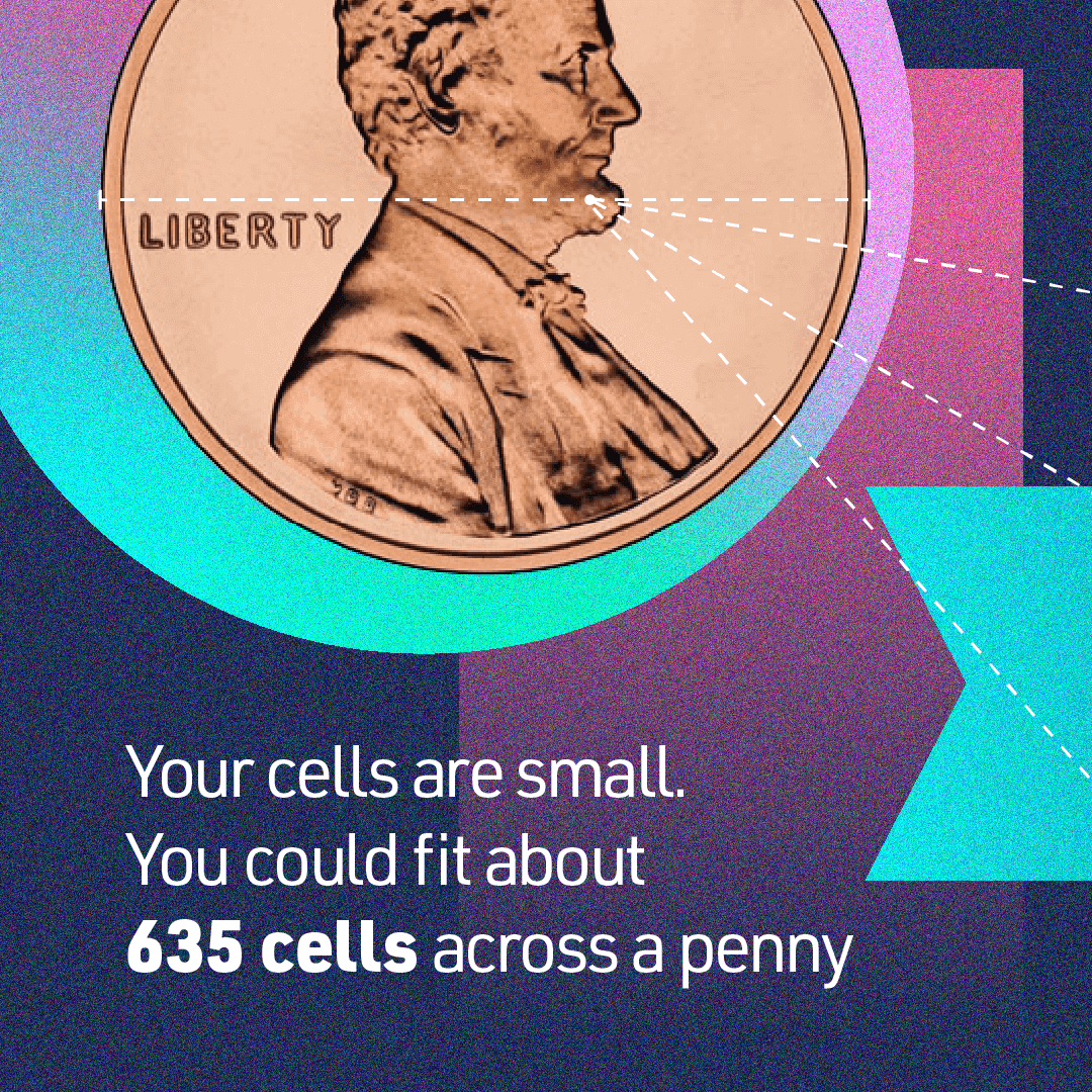
Most human cells are too small to be seen with the naked eye. For example, while a grain of salt can typically be seen without any magnification, imagine cutting it into five smaller pieces. Each of those pieces would be about the size of the average human cell. In fact, our cells are so tiny that, on average, about 635 cells could fit across the diameter of a penny.
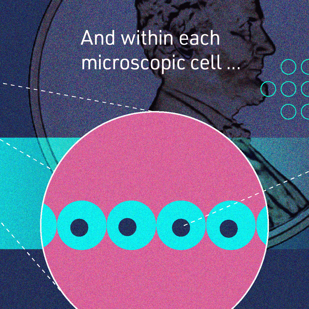
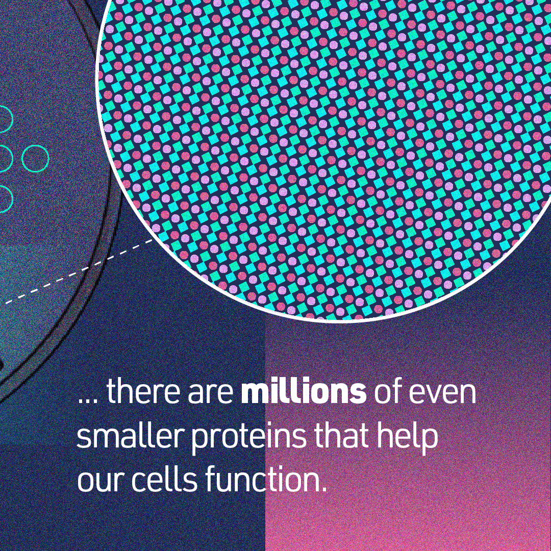
The molecular components of our cells and all of the biochemistry surrounding them are even smaller, and it’s at this molecular scale that insights to what makes our cells healthy or sick are hiding. For example, take that same grain of salt, but this time cut it into 100,000 pieces. Now we’re dealing with pieces so small, they’re measured in nanometers. That’s the scale of larger proteins you’d find within a cell that are critical for a cell’s function.
Since the 16th century, microscopes have been helping scientists get a closer look at our biology. In the last few decades, advanced imaging technologies have illuminated microscopic details about our cells. Researchers continue to push the boundaries of current technology to get a front-row seat to how these building blocks of life function in everyday health and disease.
Imaging technology doesn’t stop at the microscope. A lot of key insights gleaned from high-resolution images come from the way those images are analyzed. And imaging at the nanoscale brings new challenges to that process. For example, imaging just one cell at nanometer resolution takes up the same amount of data as about 83,000 photos on your phone.
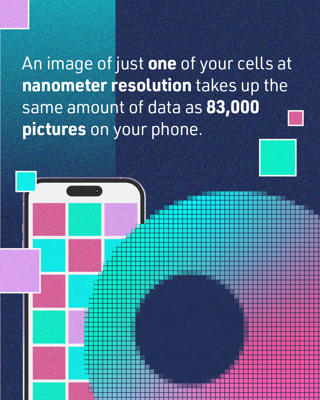
How much data is generated by imaging biology at nanometer resolution?
The research community is overcoming this data challenge with collaboration. For example, napari is a community-built, open-source tool designed for browsing, annotating and analyzing these types of large and complex images. With the CZI project napari hub, plugins created by the scientific community are openly accessible for those who want to perform advanced image analysis without any coding experience. In the last two years, researchers have downloaded napari plugins over 123,000 times.
Learn more about some of the amazing biology captured using napari.
The imaging community continues to push the limits for what is possible to observe. But with so much to discover, global collaboration is key to unlocking the next big breakthroughs.
Meet some of the imaging scientists building expert imaging networks around the world.
A ‘Google Earth’ of embryology, CZ Biohub San Francisco's zebrafish cell atlas brings a new vision to developmental biology
Learn More
Using frogs as a model organism, CZ Biohub SF Investigator Helen Willsey is revealing surprising connections between autism and hair-like structures on the...
Learn More
Watch how Jeremy Corrigan is using Chan Zuckerberg GEN EPI to track and stop outbreaks across his community.
Learn More
Stay up-to-date on the latest news, publications, competitions, and stories from CZ Biohub.
Cookies and JavaScript are required to access this form.